Ammabel’s bug blog

Ammabel Tukiman’s URAP experience: Who’s in there?

Imagine this: You’re humming a little tune to yourself as you walk by the fossil of the T-Rex in the Valley Life Sciences Building, and suddenly, you feel a little something on your skin. You look at your arm… and there’s an ant! What would you do?

If I were my previous self, I would have screamed and slapped at my arm furiously, trying to get rid of it. As a kid, I had an innate dislike for insects. The thought and sight of them always made my skin crawl, and I would always hide behind my father and cry at him to dispose of them. As I got older, this loathing I had for insects continued on. However (and thankfully), a biology course I took in community college changed the way I viewed these little – and sometimes not so little – creatures.

Despite how taxing it was to remember the taxonomy, I truly enjoyed taking the course. Having seen so many pictures of different animals, even the insects started to look adorable and interesting to me! They were no longer these disgusting creatures I found the urge to kill every single time. So, if someone were to ask the current me what I’d do if I saw an ant, or any insect really, on my arm, I would still scream but for a different reason – happiness and excitement. (Moreover, if it weren’t for my aversion to selfies, I would even take a selfie with it, before moving it to another surface.) Insects are so interesting to look at and observe – and it’s so much better to have found joy in them, instead of fear and disgust.

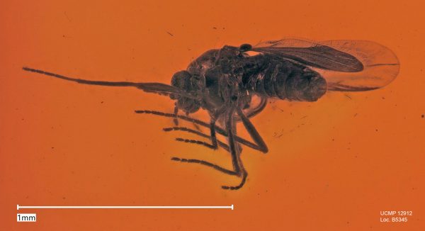
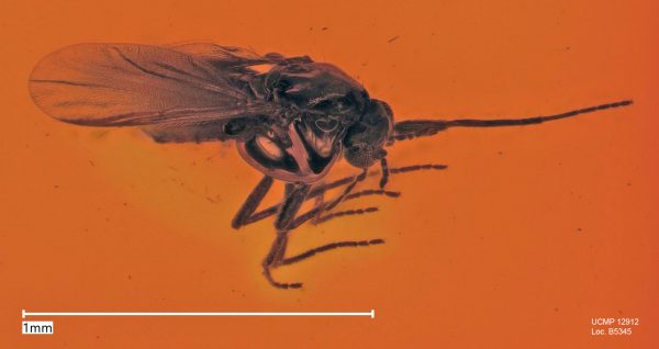
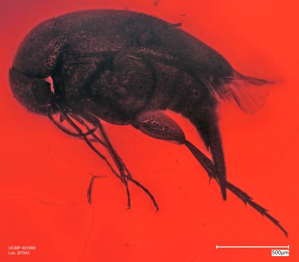
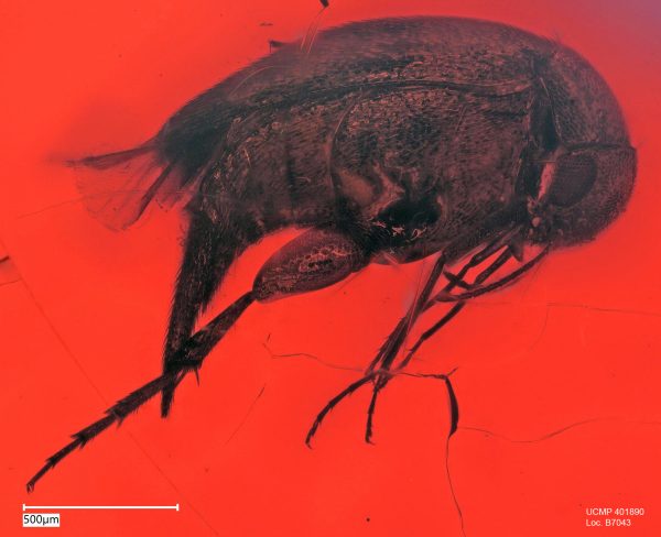
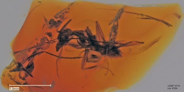
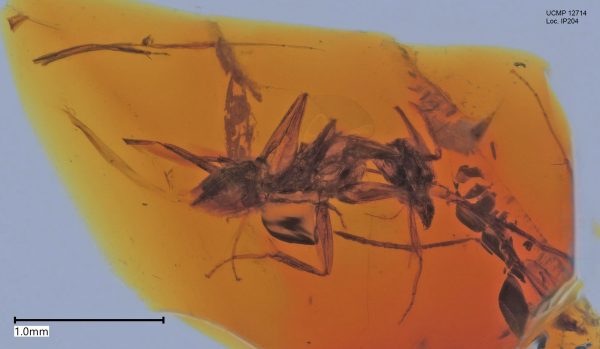
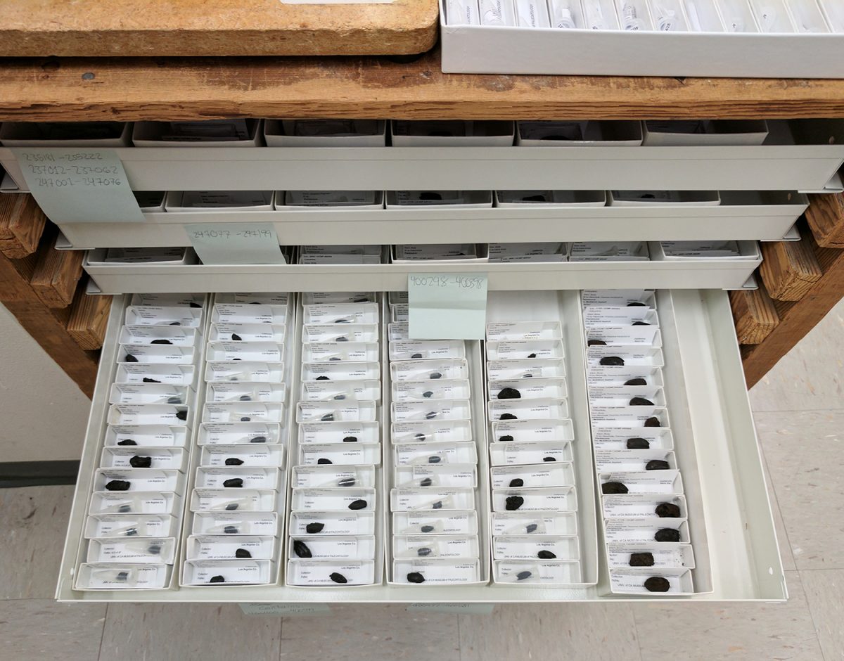
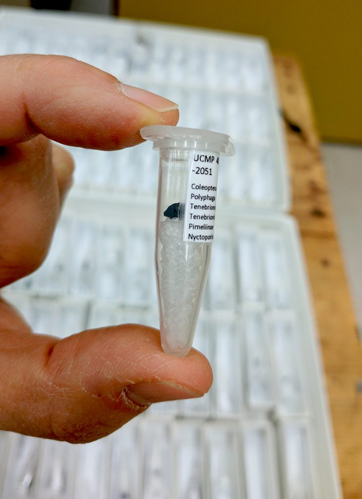
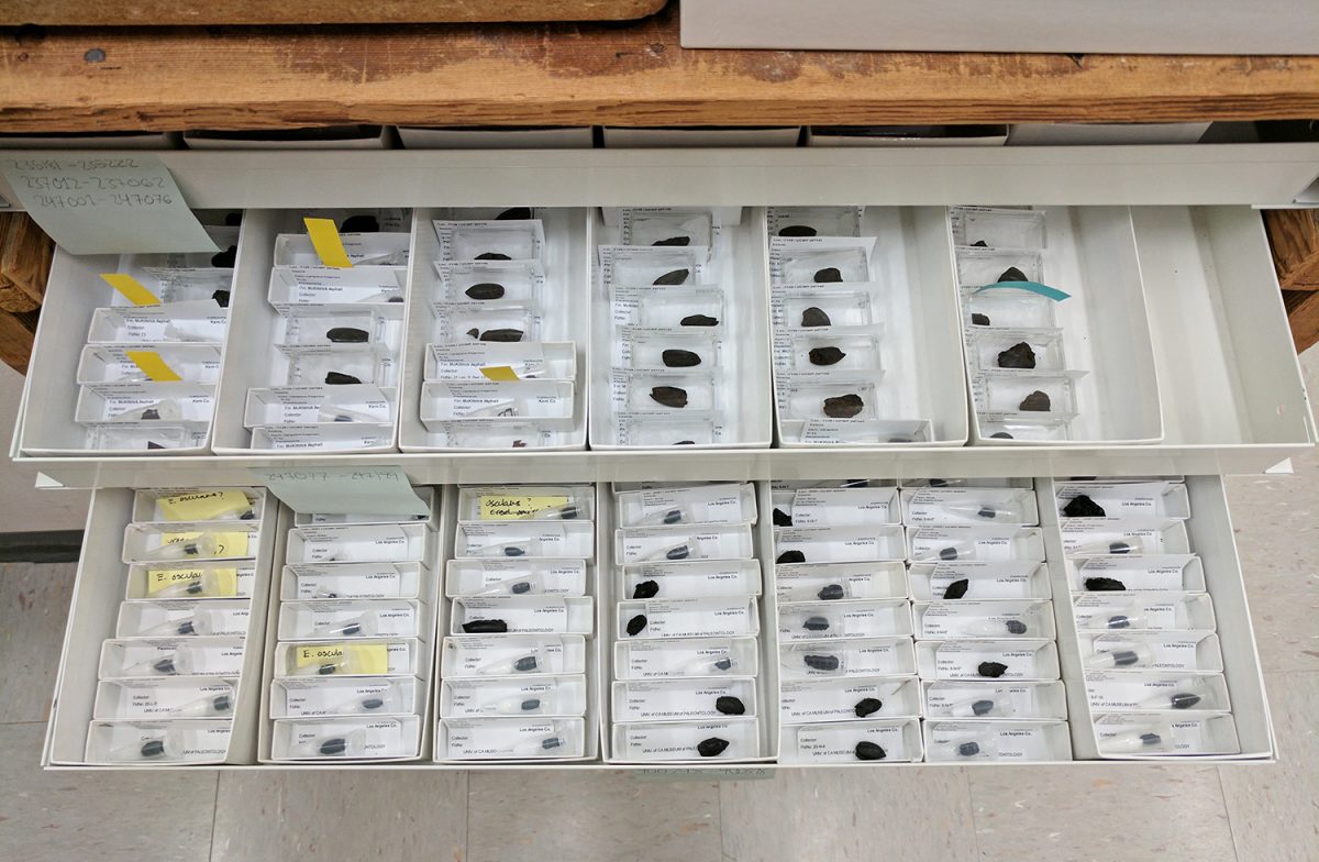
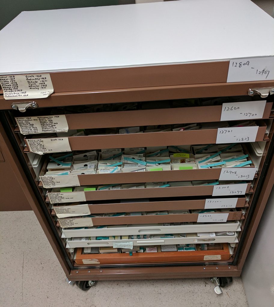
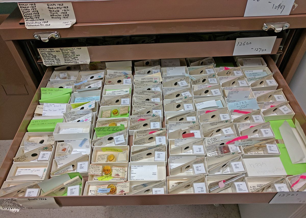
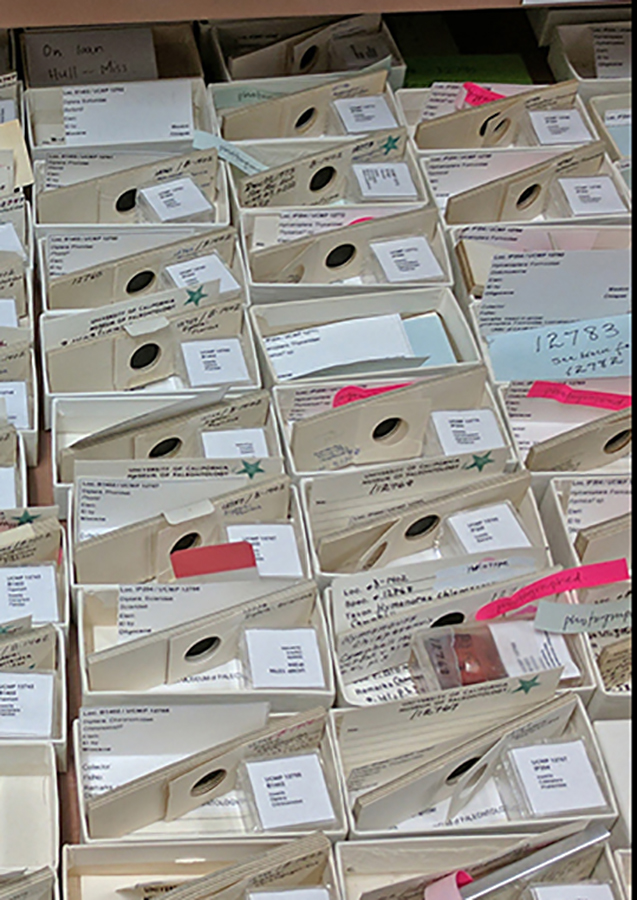
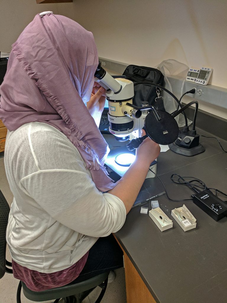
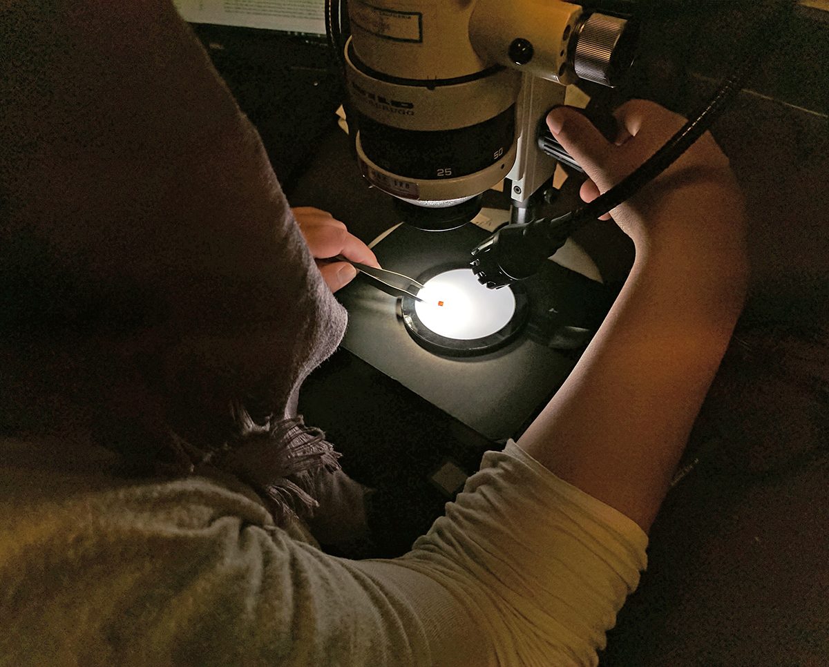
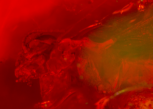
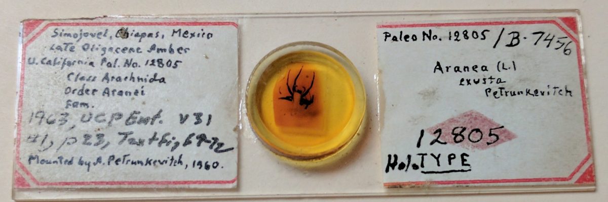
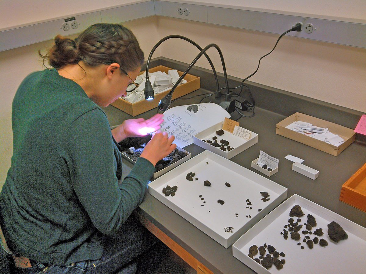
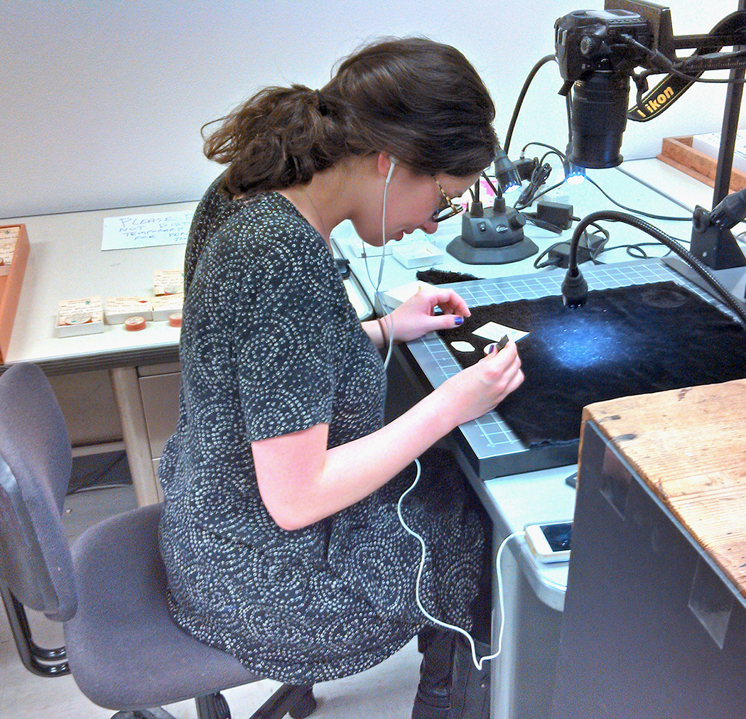
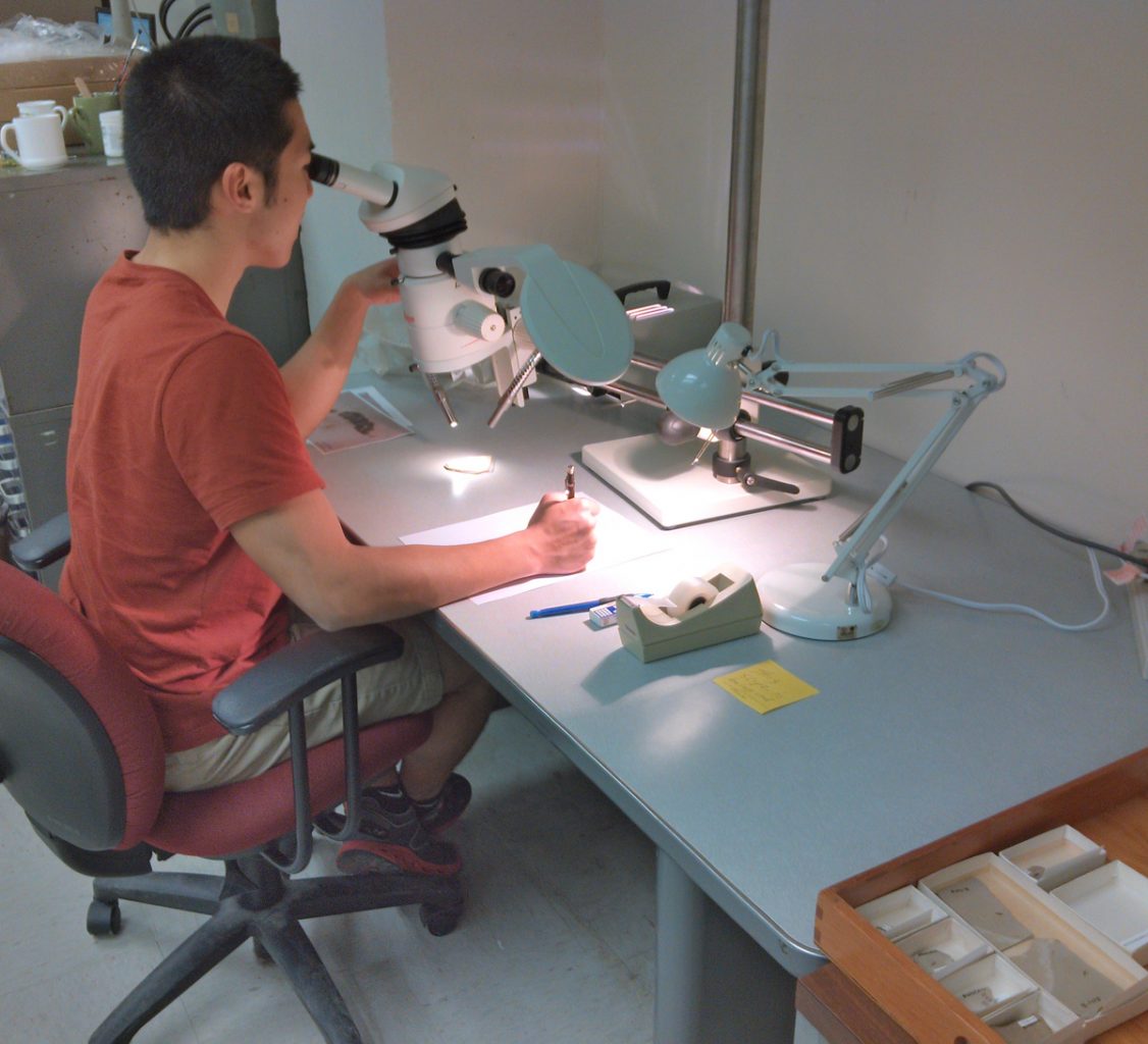
Thus, this Fall 2022 semester, I am truly honored to have gotten the opportunity to help the UC Museum of Paleontology image and digitize their amber fossil collection from Chiapas, Mexico. (More information about the UCMP Amber Collection can be found here.) Taken under the wing of Senior Museum Scientist Diane Erwin and a previous URAP student named John, I learned how to use the Keyence VHX 7000 digital microscope to image the insect inclusions inside the amber specimens. Then, I learned how to use Adobe Photoshop to process the photos, before uploading them onto CalPhotos for both researchers and interested members of the public to enjoy.

Using the Keyence VHX 7000

I’ll start off by saying that I had a very limited experience with microscopes. Before this opportunity, the only microscope I had used was one that came in a kid’s biology kit. Using that microscope was a fun experience, still, but I had my doubts about how quickly I’d be able to learn the techniques of using the Keyence VHX 7000 digital microscope with just that knowledge. And this was why I was surprised when I discovered the simplicity of such a complex instrument! 

In just the few steps written below (credited to John Feng), an image of the insect inclusions in an amber specimen can be taken.

  1. Turn on the microscope, raise the stage, and adjust the white balance.
  2. Adjust settings to high resolution mode, and ensure that full ring light and transmitted lighting are on.
  • High resolution mode gives a better image quality, while the full ring light and transmitted lighting help to illuminate the amber specimens so that the insect inclusions can be more clearly seen.

      3. Load the specimen onto the stage and find the insect inclusion(s).

  • Oftentimes, this takes a huge time of the process, especially when they are really small and there are other artifacts and debris inside the amber.

      4. Adjust the brightness, lighting, lens angle, and magnification accordingly.

  • If the specimen is not flat, we tilt the lens to make it parallel to the surface of the epoxy resin surrounding the specimen. This allows for less refraction of light, which would often result in a blurry image.
  • Usually, we go for a 100x or 200x magnification because anything lower than that would give a more noisy image. However, for bigger inclusions, a lower magnification would need to suffice.

       5. Focus up and down through the inclusion to determine the highest and lowest focal planes.

  • The microscope automatically stacks a series of consecutive images into one composite image so a critical step is determining the insect body part that is at the lowest point and focusing on that part (ie., the tip of a leg or antenna, the edge of a wing) or a little beyond it, and also the body part that is at the highest point so you know when to stop the automated imaging process, capturing the entire insect in focus.

       6. Name and save the image, and we’re done! 

  • We save the image as a JPEG file, and the naming convention we use includes the specimen number, the locality number, the date the photograph was taken, and the initials of the photographer. This aids in the processing and uploading processes.

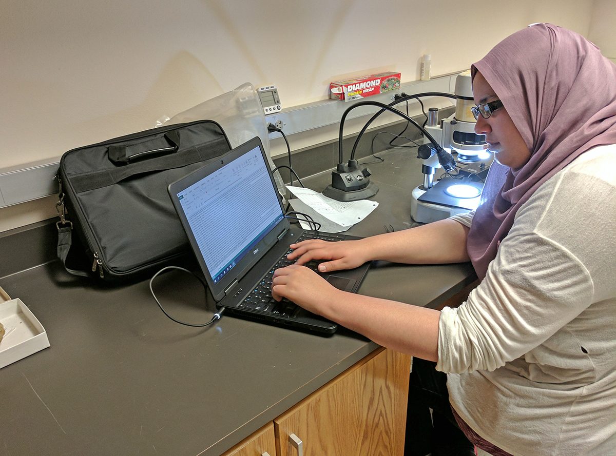
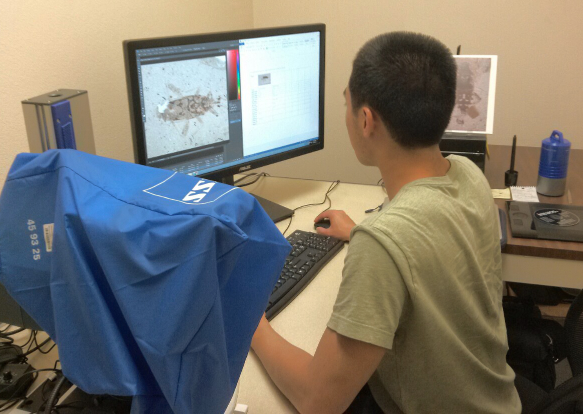
Once done with the imaging, we move on to processing the images using Adobe Photoshop.

Processing the Images

While the images taken using the Keyence VHX 7000 digital microscope are indeed clear and beautiful, they aren’t ready enough to be uploaded to CalPhotos – and that’s where Photoshop comes in. The images become even more detailed and crisp after processing!

Here’s a peek into how we process the images.

  1. Open the image file in Photoshop.
  2. Crop the image, ensuring that all parts of the insect are included.
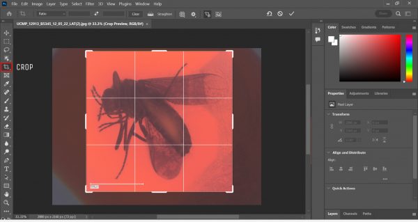
    Using the Crop function in Photoshop.
     
  3. Go to Image > Auto Color. Then, go to Image > Adjustments > Shadows/Highlights. This is where the magic happens!
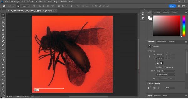
    Using Auto Color in Photoshop to enhance the contrasts in the image.

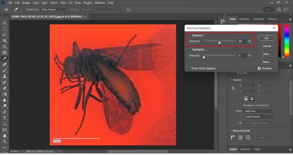
    Using Shadows/Highlights in Photoshop. Usually, we only toggle with Shadows. This makes the details of the insect more visible!
  4. Make a text box, type in the specimen number and the locality number, move the text box to a corner, and flatten the image.
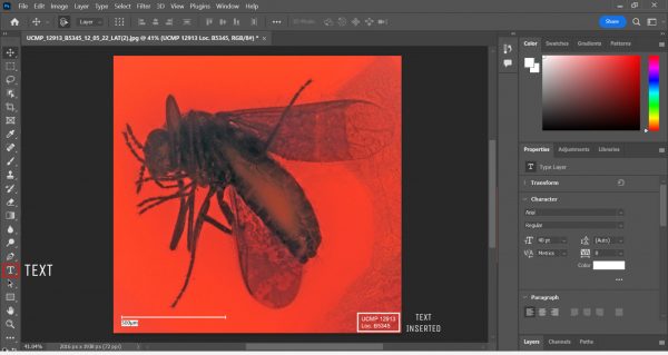
    Using the Text function in Photoshop.
  5. Save the processed image.
Here’s what the image looks like before and after processing.

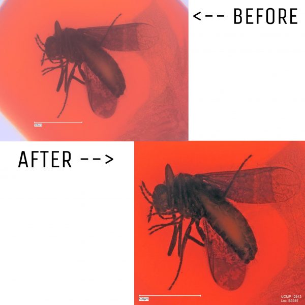
Sometimes though, in Step 3, the magic fails to happen and we end up with a whitened effect on the image. When that happens, we go to Exposure and play around with the effects to obtain the best-processed image we can make.

When we’re done with the processing, we move on to the final step – uploading the images.

Uploading the Images

I found this to be the simplest part of my URAP experience, but it was by no means any less satisfying or worthwhile. A CalPhotos contributor account was created for me, and then, I was off to upload the images that I had taken and processed.

To upload the images, I had to input only a few things – the specimen collection (in this case, it’s the Invertebrates (I) collection), the specimen number, the date the photograph was taken, the project, and the equipment used to take the image. The other details of the specimen would already be included.

The first time I uploaded my first image, I felt a huge sense of fulfillment. A particular comment from my friend, after I showed her some of my images, stuck with me. She said that I was now immortalized since my name was credited as the photographer – and it made me even more thankful that I had been given this opportunity.

I am really thankful to have gotten the chance to work together with Senior Museum Scientist Diane Erwin and help the UCMP digitize their amber fossil collection! Through my URAP experience, not only did I learn new techniques and tools while cultivating an interest of mine, but I also met some brilliant people who often made my day. It was an immense honor and pleasure to have been a part of this project!

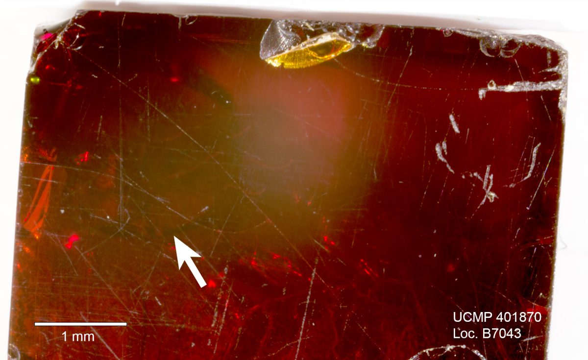
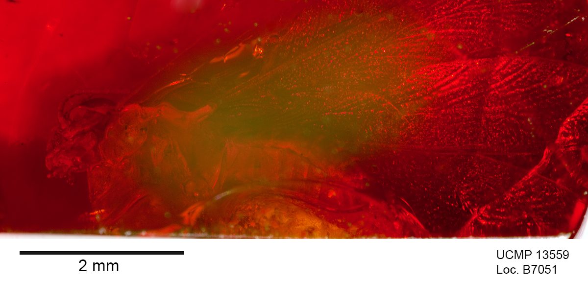
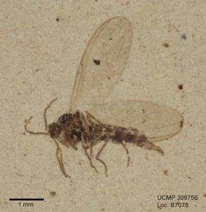
To end this, I would like to share some of my favorite images.

This insect is part of the Family Chironomidae. They resemble and are often confused for mosquitoes, but none of the members of this family are blood-feeders, which gives their common name “non-biting midges” reason.
A picture showing the other side of the above insect.
This tiny insect belongs to the Family Mordellidae, more commonly known as the tumbling flower beetles. As their name suggests, these insects tumble when escaping predators — or when they are perturbed by a human seeking to record a video of their movement.
A picture showing the other side of the above insect.
The remains of just one of many ants (Family Formicidae) preserved in the Chiapas amber. Ants have two stomachs — one for holding food for their own consumption and another for holding food to share with other ants.
A picture showing the other side of the above insect.

Madeline’s bug blog

Madeline Little’s URAP experience: A semester spent “plugging away” rehousing the UCMP tar pit collections!

Some of the type specimens collected and described in the early 1900s from the Rancho La Brea (Rosemary, CA) tar pits in their original storage units.
Digital image of the hypotype specimen UCMP 10017 published as Coniontis puncticollis LeConte (darkling beetle) by Fordyce Grinnell, Jr. in 1908, associated with its label data.

During the Spring 2017 semester, I worked as an undergraduate research apprentice at the UCMP on the Berkeley Fossil Insect PEN project to organize and rehouse the digitized insect specimens from the Rancho La Brea and McKittrick tar pits. The UCMP McKittrick collection includes insect specimens collected as early as the 1930s, while the Rancho La Brea collection has insects dating from the late 1900s when the site was first referred to as Rosemary. At this writing, over 1,300 tar pit specimens have been digitized, with images and specimen records available in CalPhotos, the UCMP online database, iDigBio, iDigPaleo, and other data aggregators. These specimens included not only those digitized during the BFIP project, but also additional ones added this year as a direct result of BFIP’s digitized data being used by Anna Holden, a doctoral student at the Richard Gilder Graduate School of the American Museum of Natural History for her dissertation research on the paleoclimate of southern California since the last Ice Age. 

During the earlier databasing and imaging stages of the project, individual insect specimens were sorted and placed in 1 x 3 inch specimen trays. Specimens curated under a previous grant had been placed in glass vials with plastic snap caps. However, because of the small size of the specimens, permanently storing them in the trays and vials would be a huge waste of space and potentially detrimental to the specimens. Maximizing the efficient use of space is always a high priority for museum collections. Storage in the trays would also leave them open to the air to collect dust, it would allow movement that could cause damage (breakage) and in the case of very small specimens or fragments, they could be blown away. I did find one of the trays was empty and learned it was a tiny specimen that was lost due to a sneeze. Additionally, I discovered specimens, which were broken or fragmented due to this storage method.

Metal drawers with labeled specimens arranged in numerical order and showing storage of 1 X 3 specimen trays. Some specimens have been rehoused into PCR tubes, the larger specimens await placement in the friction lid plastic boxes.

My first task was to organize the collection, arranging it in numerical order by specimen number. I then had to transfer the specimens into their new space-saving storage units. As part of this, I had to be sure to include all of the useful pieces of the damaged specimens and keep the specimens associated with their labels. The smaller specimens were rehoused in PCR tubes (plastic tubes with snap caps used in the field of molecular biology) with ethofoam plugs at the bottom to minimize the “rattle space,” a term coined by David Zelagin (Digitization Assistant at CU Boulder Natural History Museum). The PCR tubes would not only prevent further damage but also take up less space than the open trays.

Close-up of a specimen in their labeled PCR tube with ethofoam plug.

After labeling, they were stored upright in museum trays using the cardboard separator inserts that come as part of the packaging for the glass vials. The larger specimens were placed in small friction lid plastic specimen boxes, with archival tissue paper at the bottom to minimize rattle space and storage space. I was able to complete reorganizing of the entire

Specimen trays fitted with glass vial cardboard separator inserts to house the tar pit specimens rehoused in PCR tubes.
Specimens organized in numerical order and in various stages of rehousing. Larger specimens in the clear rectangular friction lid plastic boxes lined with tissue paper are visible in the upper drawer.

collection and rehousing all of the small and many of the larger specimens for ease of storage.

Though not the largest fossil insect collection, UCMP’s Cenozoic insects have contributed to the Fossil Insect Collaborative TCN by filling in geographic and temporal gaps not covered by the other TCN collections. I have enjoyed the opportunity to be part of the larger effort to organize and digitize this important collection so it can be accessed in an online database and more effectively used by researchers as well as the public.

Asma’s bug blog

Asma Ahmed’s URAP experience: Assessing the UCMP Amber Type specimen collection! 

I have been working the last two semesters in the UC Berkeley Museum of Paleontology on assessing the condition of the Type amber collection as part of my project for the Undergraduate Research Apprentice Program. This collection was amassed and actively published on during the 1950s to early 1970s by UC Berkeley faculty, their students and colleagues from around the world.

Long-term exposure to light can cause amber to darken so as a precaution we moved the amber collection into a light-tight Delta Designs transfer case (door removed) during the imaging, assessment and rehousing phases, which also helped provide easy access.

Together with PI Dr. Diane Erwin, I devised a system for characterizing the condition of this collection. The substandard storage of the amber specimens did not fully come to light until the digitization work started. As we discovered, the amber specimens were stored in 1 X 3 inch cardboard slide mounts sealed with a thin plastic cover slip.

Drawer of specimens showing the old cardboard slides and the new storage in labeled gem boxes.
Close-up of old slide mounts with cover slips cut out and the new labeled plastic gem boxes.

Back in the day, the cardboard slides were thought to be an adequate storage method. However, in many cases the cardboard has yellowed, indicating they are not acid free, and the cover slips were placed (forced in many cases) over specimens that were taller than the slide well. I noticed that a lot of the amber damage was centered on the fracturing that resulted from being constrained in the slides from the cover slips pressing down on the amber itself. Since the amber is very fragile, any pressure can result in multiple fractures, and the closer those fractures were to the inclusions, the less likely one can actually see the insect clearly.

Furthermore, amber pieces showed scratches on their surfaces that, in part, were likely caused by the drag of the cover slip over the specimen (and the specimen against the well bottom) when the cover slips were pulled out. Some specimens were placed in double-thick cardboard slides and these fared better because the cover slips were less likely to be pushing down on the amber.

As part of my workflow, I removed the specimens, examined them with the microscope to assess their condition, recorded my findings in an excel spreadsheet, and then transferred the specimens into ethafoam-lined, light and airtight labeled plastic gem boxes to avoid further damage. To remove the amber specimens I used a scalpel to cut one side of the cardboard so that the cover slip could be removed by lifting it up rather than sliding it out.

This way, there was no further detrimental contact between the cover slip and the specimen. There are instances where handling the amber can result in more crumbling, so all the amber must be handled very carefully when taken out of and/or placed back into housing. In handling the amber it is best to use soft forceps, which minimize stress when you squeeze them because they give in response to pressure and don’t do any damage to the amber. There are even instances where placing the amber in the new box can be damaging, so the ethafoam padding needs to be cut so that it holds the amber securely, but also so that the amber isn’t so secure it is being squeezed. In order to make sure that is the case, I would hold the amber over the ethafoam and cut a hole that is slightly larger than the amber itself. That way, the amber will stay within the hole, but also has some wiggle room that avoids any stress to the amber. 

Asma assessing fossil amber inclusions using the microscope.

Although amber darkness did impact the quality of the images, I found that in many cases it did not affect visualization of the insect unless it was coupled with extensive fracturing. Otherwise, it only blurred the image of the fossil, but didn’t make it impossible to see details. The amber specimens embedded in resin or in resin and mounted on glass microscope slides were in excellent condition: there was nothing to distract you from the fossil and you could pinpoint the location of the insect almost immediately. Fossils with internal fracturing often made shapes that look like it could be an insect, so you could mistake those patterns as an insect. Oxidized amber is generally more fragile, and therefore more likely to get fractured. All the amber has been placed in these airtight boxes and cushioned by ethafoam so that they do not move around in the boxes and undergo further damage. 

Asma using soft forceps and a combination of LED gooseneck lamps for angled side lighting and a light disc for bottom lighting.


I also noticed amber that was originally housed in the airtight gem boxes were generally in better condition than the ones that were in the cardboard slides. They were generally lighter-colored and didn’t feel that fragile when being handled by the soft forceps. This is probably due to the fact that when they are placed in airtight places, exposure to air is minimized.

Asma entering her amber assessment observations/data into the Excel spreadsheet.
Dark fractured piece of amber with scratches on surface. Inclusion at arrow.
A termite in piece of dark amber. Despite the dark nature of the amber, the inclusion is clearly visible in this image and even more so when viewed with the microscope.
Image showing detail of the head and prothorax of the termite shown above.

I assessed the amber specimens by examining their relative amount of fracturing and translucence. Amber specimens were assessed through the F-factor, a score from 1-10 that assesses how much of the amber is covered in fractures, and clarity, which is also scored on a scale from 1-10 based on how clear the amber is. There were situations where there is a lot of fracturing and/or the amber is very dark, but the area around the fossil is clear. In these cases, I made a note in the specimen comments section that the fossil is visible.

Screenshot of my spreadsheet listing the specimen number, locality number, clarity, F-factor, photograph (yes/no), embed (yes/no), and additional comments.

There were some cases where the amber specimens were given a high score for clarity (meaning it is completely dark), but the insect itself can be seen clearly. In those cases, the darkness of the amber provided the sharp contrast needed in order to see the insect itself. In the cases where the amber is very dark, it is generally too hard to see the insect at first. However, once you have a dim light shining beneath the amber, which makes the amber itself a lighter color, and no lights shining on top (because the result is a mirror effect where you can’t see anything but the glare of the light) the insect-which is very dark-becomes a stark contrast to the dark red of the amber. This allows for the clear visualization of the insect fossil, which would not have been visible beforehand.

Holotype amber specimen embedded in resin and affixed to a glass microscope slide nearly 60 years ago.

In general, the damage done to the amber has more to do with its F-factor than with clarity. So when calculating the percentage of amber that is damaged, the highly damaged ones are given an F-factor score of 5 or higher, whereas the less damaged ones are given a score of 4. At the time this blog was written, I had assessed 175 amber specimens. Of those fossils, 53 were highly damaged (30.3%) and 27 were moderately damaged (15.4%). My results show that among this sample 80 specimens will be given the highest priority for embedding in resin (45.7%). Embedding the fractured amber in resin will seal the cracks making the amber strong enough to withstand normal handling. The resin will also seal the amber from future oxidation, enhance the photographic quality and their detailed study with the microscope. 


Meschelle’s bug blog

Meschelle Thatcher’s UCMP undergraduate research experience: Beetles in Brea! 

meschelle_mckittrick_web1
Meschelle at work sorting through the bulk material of fossil insects from Rancho La Brea and McKittrick tarpits.

As an English major, I didn’t really know what to expect when I first started my URAP (Undergraduate Research Apprenticeship Program) appointment for the UC Museum of Paleontology Fossil Insect PEN (Partner to an Existing Network) funded by the National Science Foundation. All I knew was that I’d be handling fossils, and that struck the scientific chord in my imagination in perfect harmony. 

ucmp_blog_meschelle_beetles_web
Beetle specimens from the McKittrick asphalt sorted by Meschelle.

The Pleistocene Rancho La Brea tar pits in southern California are best known for their extinct exotic animals. However, I’ve learned there is more life in these asphalt seeps than saber-toothed cats, dire wolves, sloths, mastodons and camels. As part of my URAP experience I’ve been sorting the remains of beetles from the asphalt seeps at Rancho La Brea in Los Angeles and those near McKittrick, CA, in the southern San Joaquin Valley. Again, coming from a Humanities-oriented framework, I was slightly overwhelmed when my supervisor, Senior Museum Scientist, Diane Erwin, told me to pull out all the beetles I could find from bulk samples of asphalt recently discovered in the collection that had not yet been catalogued. Surely I was capable of spotting beetles, but I found myself wondering if I should also put aside things that look like legs, fragments of elytra and other beetle body parts? What if these beetle remnants should somehow alter the course of science forever? Ultimately, I found out that these anatomical fragments were indeed cute, but it wasn’t necessary to catalog them as individual specimens. Although not deserving of their own photo shoot, I learned they would be gathered for a group photo.

Fast forward to this lovely week in April, and I now have plenty of whole beetles really worth looking over. While most of them were easy to spot with the naked eye, I sometimes had to use a 10x magnifying glass to find the really small ones mixed in with the asphalt—the struggle with which I am positive any real paleontologist will identify. Undoubtedly, analyzing these fossils via zoomed in images of them will be particularly helpful. After all, the naked eye can only see so much. And when some of the beetle remains are just a few millimeters in size, we wholeheartedly welcome technology to swoop in and save the day…so long as we get the credit for our discoveries.

meschelle_type_imaging_web
Meschelle preparing Rancho La Brea type specimens for imaging with the D80 Nikon camera.

I have also helped with the imaging of the UCMP type specimens from Rancho La Brea and uploaded several dozen photos of the McKittrick beetles in the effort to digitize the diverse fossil insect collection at UCMP. By doing so, researchers, teachers, students, as well as citizen scientists and interested public all over the world will literally have accessible data at their fingertips to study. The Rancho La Brea type specimens, described and illustrated over 100 years ago, are a wonderful example of how the digitization age is allowing us to see these old collections in a new light.

ucmp_blog_meschelle_type
Type specimens of Elodes laticollis Le Conte from Rancho La Brea imaged by Meschelle.


As I move forward in life, such experiences will continue for me as a lifelong learner with interdisciplinary interests.

Hiep’s bug blog

Hiep Nguyen’s UCMP undergraduate research experience: “Scentless in Nevada”

I have been working this past year on making ‘sense” out of a fossil “scentless” plant bug from the middle Miocene Stewart Valley locality in west-central Nevada. Together with Senior Museum Scientist Diane M. Erwin we have identified a new fossil species of scentless plant bug (family Rhopalidae) from a Miocene lake bed deposit in Stewart Valley, Nevada. The study developed as a result of my participation as an Undergraduate Research Apprentice (URAP) in UC Berkeley’s fossil insect digitization PEN project (BFIP) funded by the National Science Foundation. The BFIP project is part of the Fossil Insect Collaborative Thematic Collections Network, a group of seven institutions that house our nation’s largest fossil insect collections. Their charge is to database and image these collections for public access online through the iDigBio and iDigPaleo web portals.

Hiep_3_blog
Hiep busy measuring specimens of the new Stewart Valley fossil rhopalid using BFIP images and computer-based analytical software.
Hiep_blog3
Hiep using the microscope camera-lucida setup to make tracings of his specimens to be used for his publication illustrations.

The Stewart Valley rhopalid is brown-bodied, 6.5-7.5 mm long from head to the tip of the abdomen, has numerous dark spots covering the legs, and the femur of its two back legs is noticeably wider than the others, but shows no evidence of spines. The feature that stands out most however is the striking set of dorsal markings on the insect’s abdomen. In the pictures below, you will notice a figure-8 shaped crest accompanied by four similarly-sized semicircular spots beneath it.

Diane and I worked through a long process of examining species sharing similar characteristics to our specimens. We collaborated with the Essig Museum of Entomology to examine modern counterparts to our fossil and were able to narrow the family down to the Rhopalidae. We then consulted the published literature on rhopalids and used the combination of abdominal markings and other characters to differentiate between species within Rhopalidae. What we found was that the fossil shares a number of its characters with species in several genera, but is closest to those in the subfamily Rhopalinae, tribe Rhopalini. Of especial note is the fossil’s abdominal markings, which are very reminiscent of those on the purported introduced European species, Brachycarenus tigrinus.

I wrapped up my URAP with a poster presentation for the 2016 Geological Society of America meeting held in Denver, CO detailing our findings about this newly discovered fossil insect and its evolutionary, biogeographic and paleoenvironmental implications as well as a first draft of a paper to be published on these results. The Nevada landscape of today with its miles of treeless expanse, dry lakebeds (playas), hot summers and cold winters was quite the opposite during the Miocene. Unlike today, Stewart Valley boasted an abundance of rain. A lush forest of dicotyledonous trees and nearby grasslands sporting an array of herbaceous plants surrounded a large lake teeming with aquatic life, its waters sustaining a diverse vertebrate fauna. Indeed 14.5 million years ago the Stewart Valley was an ideal habitat for scentless plant bugs to thrive and diversify.

Bringing UCMP’s Fossil Insects to Light and More!

Bringing UCMP’s Fossil Insects to Light and More!
NSF_web
The National Science Foundation.

Thanks to a two-year collections digitization grant awarded to the University of California Museum of Paleontology (UCMP) by the National Science Foundation’s, “Advancing the Digitization of Biological Collections (ADBC)” program, UC Berkeley joined the Fossil Insect Collaborative Thematic Collections Network (FIC) as a “Partner to an Existing Network (PEN)” in the summer of 2015.

FIC_web

The FIC is a consortium of seven institutions that house the largest fossil insect collections in the country. Members include the University of Colorado (Boulder), American Museum of Natural History, Harvard, Yale, University of Kansas, Illinois Natural History Survey (University of Illinois Urbana-Champaign), and the Virginia Museum of Natural History. The specimen data and images generated by these institutions go to the iDigBio, GBIFiDigPaleo and EOL web portals for online access by anyone. The BFIP data is also accessible through the UCMP and Calphotos online databases.

The BFIP project is contributing specimen data and images of its smaller, yet significant collections of insects preserved in amber from Mexico, those from the Rancho La Brea and McKittrick tar pits of southern California, and the thousands buried 14.5 million years ago in an ancient Nevada lake bed. Although the main task of the BFIP is to provide fossil insect data and images, one our BFIP team members, Dr. Marwa Wafeeq El-Faramawi became intrigued with the amber collection archives. Now one year into the project Dr. El-Faramawi has scanned the contents of nearly all 85 folders.

UCMP_B7078_399756
A beautifully preserved fossil fungus gnat from the middle Miocene of Stewart Valley, Nevada. This specimen is just one specimen among the thousands in the UCMP collection.

The amber archive files are one of the PEN project resources that we first targeted in the hope of finding clues on where some (or all) of the missing amber specimens are in order to track them down and, if lucky enough, get them back. However, going through the files (85 folders) made us realize just how important and probably highly overlooked these documents are. Basically speaking they contain all official correspondences concerning the amber and related research that Berkeley professors had with their peers all over the country and the world. They also revealed most of the specimen’s shipping details and receipts (going back and forth), in addition to drafts and manuscripts that the professors exchanged by snail mail in the old days. As official as they may seem, they definitely revealed more than that. These documents –if you read them right- tell so many stories; stories about the social / friendly or sometimes unfriendly side of the relationship between the professors and in some cases their families. Stories about the history of research in Berkeley and how it was done back then; and it may even tell you stories about the history of mail service at these times. These are the last century’s text messages and being able to view them is a one of a kind time-machine experience.

Frans_1922
A dapper young Frans Blom in 1922.

Given that most of the museum’s amber collection comes from Chiapas, Mexico, one of the most interesting folders is number 19, the Frans Blom folder, which includes his correspondences, photos, and postcards from 1952-1962. Frans Blom, also known as Pancho (as he used to sign his letters and postcards), is a highly renowned figure in the world of archaeology and the study of the Mayan civilization in Mexico and Guatemala. The great Danish archeologist and his wife the Swiss photographer Gertude Duby (Trudi) used to supply UC Berkeley Department of  Entomology’s professors Paul D. Hurd, Jr. and Ray F. Smith and UCMP’s J. Wyatt Durham with amber materials and host them in their house, the “Casa Na Bolom” or “the house of the jaguar,” during their amber collecting expeditions in Mexico. The house still existed under the management of his wife Trudi and served as a center for scholars researching Chiapas and Guatemala even after Blom died in 1963. It now serves as a museum, hotel and restaurant. His correspondence goes beyond the official in revealing his hospitality, high sense of humor and sturdy, determinate, courageous self, it also gives details about the expedition made by the UC-Berkeley research team and its before and after arrangements and complications. Frans Blom led an adventurous life that was documented in many articles and books. For more information about the great man it is definitely worth mentioning that the Bancroft Library has an invaluable genuine collection of Frans Blom’s personal and professional history that includes eleven boxes of his papers, diaries, correspondences and more from 1919 to 1942. All these items can be accessed by request for research purposes.

Follow us at weDIGBugs2 on Twitter and Instagram and the UC Museum of Paleontology Facebook page for posts relating to BFIP activities.